39 brain pictures and labels
Brain MRI: How to read MRI brain scan | Kenhub The cross-sectional cadaveric images have shown to be immensely helpful in understanding and easily tracking the relations of the brain structures on the axial MRI scans. Explore our video tutorial, quizzes, articles and labeled diagrams on this topic. Thalamus level Explore study unit Brain lobes Frontal lobe Lobus frontalis 1/5 Human brain - Wikipedia The adult human brain weighs on average about 1.2-1.4 kg (2.6-3.1 lb) which is about 2% of the total body weight, with a volume of around 1260 cm 3 in men and 1130 cm 3 in women. The cerebrum, consisting of the cerebral hemispheres, forms the largest part of the brain and overlies the other brain structures. The outer region of the hemispheres, the cerebral cortex, is grey matter ...
Mapping the brain to understand the mind - Knowable Magazine Some neuroscientists think the key to cracking that mystery is a better map of the brain's circuitry. Nearly 40 years ago, scientists achieved a milestone by completing a wiring diagram that traced all the connections of the 302 neurons of the roundworm Caenorhabditis elegans.They were traced by hand on printed sheets of electron microscope images, a meticulous and herculean task that took ...
Brain pictures and labels
Anatomy of the Brain - Simply Psychology The parietal lobe is located at the top of the brain, between the frontal and occipital lobes, above the temporal lobes (Figure 6). The parietal lobe is essential for integrating information from the body's senses in order to allow us to build a coherent picture of the world around us. Brain: Ultimate Guide to the Brain for AP® Psychology - Albert The cerebral cortex is what you picture when you think of what a brain looks like; it is the wrinkled surface of the brain that is a layer of neurons. As we grow and learn, the neurons in our cerebral cortex grow and connects with other neurons. The cerebral cortex is made up of four lobes: Parietal, Occipital, Temporal, and Frontal. New Map of Meaning in the Brain Changes Ideas About Memory | Quanta ... Because fMRI machines can measure changes in blood flow and electrical activity in the brain, neuroscientists often use them to study which parts of the cortex respond to different stimuli. Alex Huth, a neuroscientist at the University of Texas at Austin, studies how the brain represents linguistic meaning. Sasha Haagensen Photography.
Brain pictures and labels. Positions and Functions of the Four Brain Lobes - MD-Health.com The brain is divided into four sections, known as lobes (as shown in the image). The frontal lobe, occipital lobe, parietal lobe, and temporal lobe have different locations and functions that support the responses and actions of the human body. Let's start by identifying where each lobe is positioned in the brain. Position of the Lobes Three-Dimensional Convolutional Autoencoder ... - PubMed Central (PMC) Three-Dimensional Convolutional Autoencoder Extracts Features of Structural Brain Images With a "Diagnostic Label-Free" Approach: Application to Schizophrenia Datasets Hiroyuki Yamaguchi, 1,2 Yuki Hashimoto, 1 Genichi Sugihara, 3 Jun Miyata, 4 Toshiya Murai, 4 Hidehiko Takahashi, 3 Manabu Honda, 1 Akitoyo Hishimoto, 2 and Yuichi Yamashita 1,* Brain: Atlas of human anatomy with MRI - e-Anatomy - IMAIOS Anatomy of the brain (MRI) - cross-sectional atlas of human anatomy. The module on the anatomy of the brain based on MRI with axial slices was redesigned, having received multiple requests from users for coronal and sagittal slices. The elaboration of this new module, its labeling of more than 524 structures on 379 MRI images in three different ... Left Brain vs. Right Brain: Characteristics Chart [INFOGRAPHIC] Brain dominance theory is absorbing and enjoyable, plus it allows people to think about stereotypes and labels. In reality, though, psychology is complicated, and the truth is that there are very few people who have the traits of only one of these descriptions. More often, we have an intricate combination of both.
Free Printable Brain Hemisphere Hat - Homeschool Giveaways Free download printable Brain Hemisphere Hat. card stock. piece of paper. glue. scissors. tape. crayons. colored pencils. The download is available in color as well as black and white so that your child can color it in. Cerebral Cortex | Facts, Layers, Levels, Functions & Summary The cerebral cortex ( cortex cerebri) is the outer layer of our brain that has a wrinkled appearance. It is divided into fields with specific functions such as sight, hearing, smell, and sensation, and controls higher functions such as speech, thinking, and memory. The most important part of the brain related to self-development techniques is ... 14 Informative Facts, Diagram & Parts Of Human Brain For Kids The brain and spinal cord are part of the central nervous system (CNS). The brain weighs just about two to three pounds and appears like a walnut. The brain is comprised of three main regions — cerebrum, cerebellum, and brainstem (3). Save Image: Shutterstock Let us discuss these parts and their functions in more detail (1) (3) (4). Cross-sectional anatomy of the brain - e-Anatomy - IMAIOS Anatomy of the brain: how to view anatomical labels. This module is a comprehensive and affordable learning tool for medical students and residents and especially for neuroradiologists and radiation oncologists. It provides access to an atlas and to images in axial planes, allowing the user to learn and review neuroanatomy interactively.
Free Nervous System Worksheets and Printables The Human Body Systems Labeling and Diagramming Worksheet includes fill-in-the-blanks for your children to label the parts of the brain, spinal cord, ganglion, and nerves. Free Human Anatomy Coloring Pages for Students. A fun way to introduce your kids to anatomy is with coloring pages. We have listed out some human anatomy coloring pages that ... Are labels like gay, straight, bisexual more harm than good? The brief case for labels and definitions. ... put it in the search bar, and pictures of people like me would pop up. Looking at them in skirts and shining jewellery would make me smile." ... shouting some rhetoric. Now look around and see how far these 'brain images' are from reality. We have animated labels with our imagination. They ... What are the 12 cranial nerves? Functions and diagram The cranial nerves are a set of twelve nerves that originate in the brain. Each has a different function responsible for sense or movement. The functions of the cranial nerves are sensory, motor ... Brain: Function and Anatomy, Conditions, and Health Tips Some of its main functions include: processing sensory information regulating blood pressure and breathing releasing hormones Brain diagram Use this interactive 3-D diagram to explore the brain....
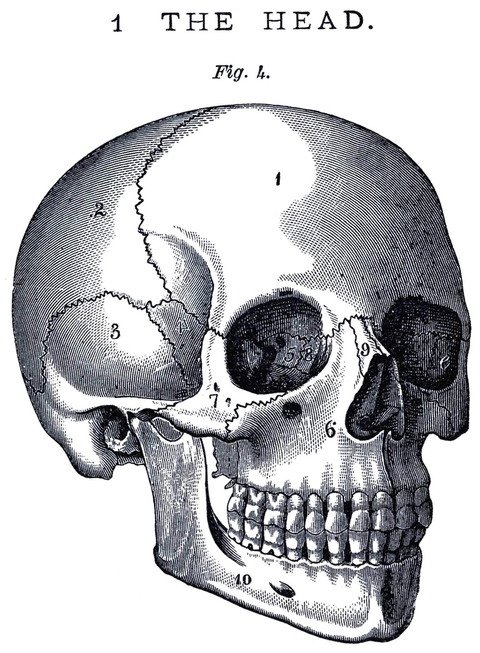
Vintage Anatomy Skull Image - The Graphics Fairy
Parts of the Brain Activity for Kids, Brain Diagram, and Worksheets for ... PARIETAL LOBES - The parietal lobe provides sensory information to the brain including touch, pain and temperature. OCCIPITAL LOBES - The occipital lobe processes and interprets everything we see TEMPORAL LOBES - The temporal lobe controls emotions and short-term memory
.jpg)
Lateral View of the Brain Centered at the Level of the Intraparietal Sulcus | Neuroanatomy | The ...
Human Brain Lesson for Kids: Function & Diagram - Study.com The Cerebrum. The biggest part of your brain is called the cerebrum. One thing your cerebrum does is control your muscles. It has two halves, one on the left and one on the right. The weird thing ...
Lateral Exposure of Middle Cerebral Artery in Sylvian Fissure | Neuroanatomy | The Neurosurgical ...
100,000 human brain images 100,000 human brain images. Jorge Cardoso, researcher in Artificial Medical Intelligence at King's College London, is heading the project. Researchers at King's College London used NVIDIA Cambridge -1, UK's most powerful supercomputer dedicated to AI research in healthcare and MONAI, an open source AI software framework, to create 100,000 ...

Brain and Spinal Cord | ClipArt ETC
Brain Anatomy: Lesson for Kids - Video & Lesson Transcript | Study.com The brain is made up of three major parts: they're called the brain stem, the cerebellum, and the cerebrum. The brain stem connects the base of the brain to the spinal cord. It's the part of the...

What are the 4 Main Types of Electrical Injury? - Pat Labels
Frontiers | Three-Dimensional Convolutional Autoencoder Extracts ... Three-Dimensional Convolutional Autoencoder Extracts Features of Structural Brain Images With a "Diagnostic Label-Free" Approach: Application to Schizophrenia Datasets Hiroyuki Yamaguchi 1,2, Yuki Hashimoto 1, Genichi Sugihara 3, Jun Miyata 4, Toshiya Murai 4, Hidehiko Takahashi 3, Manabu Honda 1, Akitoyo Hishimoto 2 and Yuichi Yamashita 1*
Cross Section of Midbrain | Neuroanatomy | The Neurosurgical Atlas, by Aaron Cohen-Gadol, M.D.
Medical Image Pre-Processing with Python | by Esma Sert | Towards Data ... brain_image = window_image (hu_image, 40, 80) #bone windowing segmentation = morphology.dilation (brain_image, np.ones ( (1, 1))) labels, label_nb = ndimage.label (segmentation) label_count = np.bincount (labels.ravel ().astype (np.int)) label_count [0] = 0 mask = labels == label_count.argmax () mask = morphology.dilation (mask, np.ones ( (1, 1)))

CNS: Ultrasound Anatomy, Brain - OB Images - OB Images
Parts of the brain: Learn with diagrams and quizzes - Kenhub Labeled brain diagram First up, have a look at the labeled brain structures on the image below. Try to memorize the name and location of each structure, then proceed to test yourself with the blank brain diagram provided below. Labeled diagram showing the main parts of the brain Blank brain diagram (free download!)

Parasagittal View of Brain and Lateral Head | Neuroanatomy | The Neurosurgical Atlas
Brain charts - University of Cambridge An international team of researchers has created a series of brain charts spanning our entire lifespan - from a 15 week old fetus to 100 year old adult - that show how our brains expand rapidly in early life and slowly shrink as we age.. The charts are the result of a research project spanning six continents and bringing together possibly the largest ever MRI datasets ever aggregated ...
Post a Comment for "39 brain pictures and labels"